partial quote from:
Microfluidics - Wikipedia
If this is true this sentence then what are DNA chips and what is lab-on-a-chip technology?
begin quote from:
DNA chips
A DNA microarray (also commonly known as DNA chip or biochip) is a collection of microscopic DNA spots attached to a solid surface. Scientists use DNA microarrays to measure the expression levels of large numbers of genes simultaneously or to genotype multiple regions of a genome. Each DNA spot contains picomoles (10−12 moles) of a specific DNA sequence, known as probes (or reporters or oligos). These can be a short section of a gene or other DNA element that are used to hybridize a cDNA or cRNA (also called anti-sense RNA) sample (called target) under high-stringency conditions. Probe-target hybridization is usually detected and quantified by detection of fluorophore-, silver-, or chemiluminescence-labeled targets to determine relative abundance of nucleic acid sequences in the target. The original nucleic acid arrays were macro arrays approximately 9 cm × 12 cm and the first computerized image based analysis was published in 1981.[1]Contents
Principle

Hybridization of the target to the probe
Main article: Nucleic acid hybridization
For more details on this topic, see § A typical protocol.
The core principle behind microarrays is hybridization between two DNA strands, the property of complementary nucleic acid sequences to specifically pair with each other by forming hydrogen bonds between complementary nucleotide base pairs. A high number of complementary base pairs in a nucleotide sequence means tighter non-covalent
bonding between the two strands. After washing off non-specific bonding
sequences, only strongly paired strands will remain hybridized.
Fluorescently labeled target sequences that bind to a probe sequence
generate a signal that depends on the hybridization conditions (such as
temperature), and washing after hybridization. Total strength of the
signal, from a spot (feature), depends upon the amount of target sample
binding to the probes present on that spot. Microarrays use relative
quantitation in which the intensity of a feature is compared to the
intensity of the same feature under a different condition, and the
identity of the feature is known by its position.
The steps required in a microarray experiment
Uses and types

Two Affymetrix chips. A match is shown at bottom left for size comparison.
- The traditional solid-phase array is a collection of orderly microscopic "spots", called features, each with thousands of identical and specific probes attached to a solid surface, such as glass, plastic or silicon biochip (commonly known as a genome chip, DNA chip or gene array). Thousands of these features can be placed in known locations on a single DNA microarray.
- The alternative bead array is a collection of microscopic polystyrene beads, each with a specific probe and a ratio of two or more dyes, which do not interfere with the fluorescent dyes used on the target sequence.
Applications include:
| Application or technology | Synopsis |
|---|---|
| Gene expression profiling | In an mRNA or gene expression profiling experiment the expression levels of thousands of genes are simultaneously monitored to study the effects of certain treatments, diseases, and developmental stages on gene expression. For example, microarray-based gene expression profiling can be used to identify genes whose expression is changed in response to pathogens or other organisms by comparing gene expression in infected to that in uninfected cells or tissues.[2] |
| Comparative genomic hybridization | Assessing genome content in different cells or closely related organisms.[3][4] |
| GeneID | Small microarrays to check IDs of organisms in food and feed (like GMO [1]), mycoplasms in cell culture, or pathogens for disease detection, mostly combining PCR and microarray technology. |
| Chromatin immunoprecipitation on Chip | DNA sequences bound to a particular protein can be isolated by immunoprecipitating that protein (ChIP), these fragments can be then hybridized to a microarray (such as a tiling array) allowing the determination of protein binding site occupancy throughout the genome. Example protein to immunoprecipitate are histone modifications (H3K27me3, H3K4me2, H3K9me3, etc.), Polycomb-group protein (PRC2:Suz12, PRC1:YY1) and trithorax-group protein (Ash1) to study the epigenetic landscape or RNA Polymerase II to study the transcription landscape. |
| DamID | Analogously to ChIP, genomic regions bound by a protein of interest can be isolated and used to probe a microarray to determine binding site occupancy. Unlike ChIP, DamID does not require antibodies but makes use of adenine methylation near the protein's binding sites to selectively amplify those regions, introduced by expressing minute amounts of protein of interest fused to bacterial DNA adenine methyltransferase. |
| SNP detection | Identifying single nucleotide polymorphism among alleles within or between populations.[5] Several applications of microarrays make use of SNP detection, including genotyping, forensic analysis, measuring predisposition to disease, identifying drug-candidates, evaluating germline mutations in individuals or somatic mutations in cancers, assessing loss of heterozygosity, or genetic linkage analysis. |
| Alternative splicing detection | An exon junction array design uses probes specific to the expected or potential splice sites of predicted exons for a gene. It is of intermediate density, or coverage, to a typical gene expression array (with 1–3 probes per gene) and a genomic tiling array (with hundreds or thousands of probes per gene). It is used to assay the expression of alternative splice forms of a gene. Exon arrays have a different design, employing probes designed to detect each individual exon for known or predicted genes, and can be used for detecting different splicing isoforms. |
| Fusion genes microarray | A Fusion gene microarray can detect fusion transcripts, e.g. from cancer specimens. The principle behind this is building on the alternative splicing microarrays. The oligo design strategy enables combined measurements of chimeric transcript junctions with exon-wise measurements of individual fusion partners. |
| Tiling array | Genome tiling arrays consist of overlapping probes designed to densely represent a genomic region of interest, sometimes as large as an entire human chromosome. The purpose is to empirically detect expression of transcripts or alternatively spliced forms which may not have been previously known or predicted. |
| Double-stranded B-DNA microarrays | Right-handed double-stranded B-DNA microarrays can be used to characterize novel drugs and biologicals that can be employed to bind specific regions of immobilized, intact, double-stranded DNA. This approach can be used to inhibit gene expression.[6][7][8] They also allow for characterization of their structure under different environmental conditions. |
| Double-stranded Z-DNA microarrays | Left-handed double-stranded Z-DNA microarrays can be used to identify short sequences of the alternative Z-DNA structure located within longer stretches of right-handed B-DNA genes (e.g., transcriptional enhancement, recombination, RNA editing).[6][7][8][9] The microarrays also allow for characterization of their structure under different environmental conditions. |
| Multi-stranded DNA microarrays (triplex-DNA microarrays and quadruplex-DNA microarrays) | Multi-stranded DNA and RNA microarrays can be used to identify novel drugs that bind to these multi-stranded nucleic acid sequences. This approach can be used to discover new drugs and biologicals that have the ability to inhibit gene expression.[6][7][8][10][11] These microarrays also allow for characterization of their structure under different environmental conditions. |
Fabrication
Microarrays can be manufactured in different ways, depending on the number of probes under examination, costs, customization requirements, and the type of scientific question being asked. Arrays may have as few as 10 probes or up to 2.1 million micrometre-scale probes from commercial vendors.Spotted vs. in situ synthesised arrays
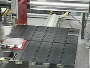
A DNA microarray being printed by a robot at the University of Delaware
In spotted microarrays, the probes are oligonucleotides, cDNA or small fragments of PCR products that correspond to mRNAs. The probes are synthesized prior to deposition on the array surface and are then "spotted" onto glass. A common approach utilizes an array of fine pins or needles controlled by a robotic arm that is dipped into wells containing DNA probes and then depositing each probe at designated locations on the array surface. The resulting "grid" of probes represents the nucleic acid profiles of the prepared probes and is ready to receive complementary cDNA or cRNA "targets" derived from experimental or clinical samples. This technique is used by research scientists around the world to produce "in-house" printed microarrays from their own labs. These arrays may be easily customized for each experiment, because researchers can choose the probes and printing locations on the arrays, synthesize the probes in their own lab (or collaborating facility), and spot the arrays. They can then generate their own labeled samples for hybridization, hybridize the samples to the array, and finally scan the arrays with their own equipment. This provides a relatively low-cost microarray that may be customized for each study, and avoids the costs of purchasing often more expensive commercial arrays that may represent vast numbers of genes that are not of interest to the investigator. Publications exist which indicate in-house spotted microarrays may not provide the same level of sensitivity compared to commercial oligonucleotide arrays,[14] possibly owing to the small batch sizes and reduced printing efficiencies when compared to industrial manufactures of oligo arrays.
In oligonucleotide microarrays, the probes are short sequences designed to match parts of the sequence of known or predicted open reading frames. Although oligonucleotide probes are often used in "spotted" microarrays, the term "oligonucleotide array" most often refers to a specific technique of manufacturing. Oligonucleotide arrays are produced by printing short oligonucleotide sequences designed to represent a single gene or family of gene splice-variants by synthesizing this sequence directly onto the array surface instead of depositing intact sequences. Sequences may be longer (60-mer probes such as the Agilent design) or shorter (25-mer probes produced by Affymetrix) depending on the desired purpose; longer probes are more specific to individual target genes, shorter probes may be spotted in higher density across the array and are cheaper to manufacture. One technique used to produce oligonucleotide arrays include photolithographic synthesis (Affymetrix) on a silica substrate where light and light-sensitive masking agents are used to "build" a sequence one nucleotide at a time across the entire array.[15] Each applicable probe is selectively "unmasked" prior to bathing the array in a solution of a single nucleotide, then a masking reaction takes place and the next set of probes are unmasked in preparation for a different nucleotide exposure. After many repetitions, the sequences of every probe become fully constructed. More recently, Maskless Array Synthesis from NimbleGen Systems has combined flexibility with large numbers of probes.[16]
Two-channel vs. one-channel detection

Diagram of typical dual-colour microarray experiment
Oligonucleotide microarrays often carry control probes designed to hybridize with RNA spike-ins. The degree of hybridization between the spike-ins and the control probes is used to normalize the hybridization measurements for the target probes. Although absolute levels of gene expression may be determined in the two-color array in rare instances, the relative differences in expression among different spots within a sample and between samples is the preferred method of data analysis for the two-color system. Examples of providers for such microarrays includes Agilent with their Dual-Mode platform, Eppendorf with their DualChip platform for colorimetric Silverquant labeling, and TeleChem International with Arrayit.
In single-channel microarrays or one-color microarrays, the arrays provide intensity data for each probe or probe set indicating a relative level of hybridization with the labeled target. However, they do not truly indicate abundance levels of a gene but rather relative abundance when compared to other samples or conditions when processed in the same experiment. Each RNA molecule encounters protocol and batch-specific bias during amplification, labeling, and hybridization phases of the experiment making comparisons between genes for the same microarray uninformative. The comparison of two conditions for the same gene requires two separate single-dye hybridizations. Several popular single-channel systems are the Affymetrix "Gene Chip", Illumina "Bead Chip", Agilent single-channel arrays, the Applied Microarrays "CodeLink" arrays, and the Eppendorf "DualChip & Silverquant". One strength of the single-dye system lies in the fact that an aberrant sample cannot affect the raw data derived from other samples, because each array chip is exposed to only one sample (as opposed to a two-color system in which a single low-quality sample may drastically impinge on overall data precision even if the other sample was of high quality). Another benefit is that data are more easily compared to arrays from different experiments as long as batch effects have been accounted for.
One channel microarray may be the only choice in some situations. Suppose
 samples need to be compared: then the number of experiments required
using the two channel arrays quickly becomes unfeasible, unless a sample
is used as a reference.
samples need to be compared: then the number of experiments required
using the two channel arrays quickly becomes unfeasible, unless a sample
is used as a reference.| number of samples | one-channel microarray | two channel microarray |
two channel microarray (with reference) |
|---|---|---|---|
| 1 | 1 | 1 | 1 |
| 2 | 2 | 1 | 1 |
| 3 | 3 | 3 | 2 |
| 4 | 4 | 6 | 3 |
|
|
 |
 |
A typical protocol
This is an example of a DNA microarray experiment, detailing a particular case to better explain DNA microarray experiments, while enumerating possible alternatives.- The two samples to be compared (pairwise comparison) are grown/acquired. In this example treated sample (case) and untreated sample (control).
- The nucleic acid of interest is purified: this can be all RNA for expression profiling, DNA for comparative hybridization, or DNA/RNA bound to a particular protein which is immunoprecipitated (ChIP-on-chip) for epigenetic or regulation studies. In this example total RNA is isolated (total as it is nuclear and cytoplasmic) by Guanidinium thiocyanate-phenol-chloroform extraction (e.g. Trizol) which isolates most RNA (whereas column methods have a cut off of 200 nucleotides) and if done correctly has a better purity.
- The purified RNA is analysed for quality (by capillary electrophoresis) and quantity (for example, by using a NanoDrop or NanoPhotometer spectrometer). If the material is of acceptable quality and sufficient quantity is present (e.g., >1μg, although the required amount varies by microarray platform), the experiment can proceed.
- The labelled product is generated via reverse transcription and sometimes with an optional PCR amplification. The RNA is reverse transcribed with either polyT primers (which amplify only mRNA) or random primers (which amplify all RNA, most of which is rRNA); miRNA microarrays ligate an oligonucleotide to the purified small RNA (isolated with a fractionator), which is then reverse transcribed and amplified. The label is added either during the reverse transcription step, or following amplification if it is performed. The sense labelling is dependent on the microarray; e.g. if the label is added with the RT mix, the cDNA is antisense and the microarray probe is sense, except in the case of negative controls. The label is typically fluorescent; only one machine uses radiolabels. The labelling can be direct (not used) or indirect (requires a coupling stage). For two-channel arrays, the coupling stage occurs before hybridization, using aminoallyl uridine triphosphate (aminoallyl-UTP, or aaUTP) and NHS amino-reactive dyes (such as cyanine dyes); for single-channel arrays, the coupling stage occurs after hybridization, using biotin and labelled streptavidin. The modified nucleotides (usually in a ratio of 1 aaUTP: 4 TTP (thymidine triphosphate)) are added enzymatically in a low ratio to normal nucleotides, typically resulting in 1 every 60 bases. The aaDNA is then purified with a column (using a phosphate buffer solution, as Tris contains amine groups). The aminoallyl group is an amine group on a long linker attached to the nucleobase, which reacts with a reactive dye. A form of replicate known as a dye flip can be performed to remove any dye effects in two-channel experiments; for a dye flip, a second slide is used, with the labels swapped (the sample that was labeled with Cy3 in the first slide is labeled with Cy5, and vice versa). In this example, aminoallyl-UTP is present in the reverse-transcribed mixture.
- The labeled samples are then mixed with a propriety hybridization solution which can consist of SDS, SSC, dextran sulfate, a blocking agent (such as COT1 DNA, salmon sperm DNA, calf thymus DNA, PolyA or PolyT), Denhardt's solution, or formamine.
- The mixture is denatured and added to the pinholes of the microarray. The holes are sealed and the microarray hybridized, either in a hyb oven, where the microarray is mixed by rotation, or in a mixer, where the microarray is mixed by alternating pressure at the pinholes.
- After an overnight hybridization, all nonspecific binding is washed off (SDS and SSC).
- The microarray is dried and scanned by a machine that uses a laser to excite the dye and measures the emission levels with a detector.
- The image is gridded with a template and the intensities of each feature (composed of several pixels) is quantified.
- The raw data is normalized; the simplest normalization method is to subtract background intensity and scale so that the total intensities of the features of the two channels are equal, or to use the intensity of a reference gene to calculate the t-value for all of the intensities. More sophisticated methods include z-ratio, loess and lowess regression and RMA (robust multichip analysis) for Affymetrix chips (single-channel, silicon chip, in situ synthesised short oligonucleotides).
Microarrays and bioinformatics

Gene expression values from microarray experiments can be represented as heat maps to visualize the result of data analysis.
- the multiple levels of replication in experimental design (Experimental design)
- the number of platforms and independent groups and data format (Standardization)
- the statistical treatment of the data (Statistical analysis)
- mapping each probe to the mRNA transcript that it measures (Annotation)
- the sheer volume of data and the ability to share it (Data warehousing)
Experimental design
Due to the biological complexity of gene expression, the considerations of experimental design that are discussed in the expression profiling article are of critical importance if statistically and biologically valid conclusions are to be drawn from the data.There are three main elements to consider when designing a microarray experiment. First, replication of the biological samples is essential for drawing conclusions from the experiment. Second, technical replicates (two RNA samples obtained from each experimental unit) help to ensure precision and allow for testing differences within treatment groups. The biological replicates include independent RNA extractions and technical replicates may be two aliquots of the same extraction. Third, spots of each cDNA clone or oligonucleotide are present as replicates (at least duplicates) on the microarray slide, to provide a measure of technical precision in each hybridization. It is critical that information about the sample preparation and handling is discussed, in order to help identify the independent units in the experiment and to avoid inflated estimates of statistical significance.[19]
Standardization
Microarray data is difficult to exchange due to the lack of standardization in platform fabrication, assay protocols, and analysis methods. This presents an interoperability problem in bioinformatics. Various grass-roots open-source projects are trying to ease the exchange and analysis of data produced with non-proprietary chips:- For example, the "Minimum Information About a Microarray Experiment" (MIAME) checklist helps define the level of detail that should exist and is being adopted by many journals as a requirement for the submission of papers incorporating microarray results. But MIAME does not describe the format for the information, so while many formats can support the MIAME requirements, as of 2007 no format permits verification of complete semantic compliance.
- The "MicroArray Quality Control (MAQC) Project" is being conducted by the US Food and Drug Administration (FDA) to develop standards and quality control metrics which will eventually allow the use of MicroArray data in drug discovery, clinical practice and regulatory decision-making.[20]
- The MGED Society has developed standards for the representation of gene expression experiment results and relevant annotations.
Data analysis
See also: Gene chip analysis
Microarray data sets are commonly very large, and analytical precision is influenced by a number of variables. Statistical challenges include taking into account effects of background noise and appropriate normalization
of the data. Normalization methods may be suited to specific platforms
and, in the case of commercial platforms, the analysis may be
proprietary.[citation needed] Algorithms that affect statistical analysis include:- Image analysis: gridding, spot recognition of the scanned image (segmentation algorithm), removal or marking of poor-quality and low-intensity features (called flagging).
- Data processing: background subtraction (based on global or local background), determination of spot intensities and intensity ratios, visualisation of data (e.g. see MA plot), and log-transformation of ratios, global or local normalization of intensity ratios, and segmentation into different copy number regions using step detection algorithms.[21]
- Class discovery analysis: This analytic approach, sometimes called
unsupervised classification or knowledge discovery, tries to identify
whether microarrays (objects, patients, mice, etc.) or genes cluster
together in groups. Identifying naturally existing groups of objects
(microarrays or genes) which cluster together can enable the discovery
of new groups that otherwise were not previously known to exist. During
knowledge discovery analysis, various unsupervised classification
techniques can be employed with DNA microarray data to identify novel
clusters (classes) of arrays.[22]
This type of approach is not hypothesis-driven, but rather is based on
iterative pattern recognition or statistical learning methods to find an
"optimal" number of clusters in the data. Examples of unsupervised
analyses methods include self-organizing maps, neural gas, k-means
cluster analyses,[23] hierarchical cluster analysis, Genomic Signal Processing based clustering[24]
and model-based cluster analysis. For some of these methods the user
also has to define a distance measure between pairs of objects. Although
the Pearson correlation coefficient is usually employed, several other
measures have been proposed and evaluated in the literature.[25]
The input data used in class discovery analyses are commonly based on
lists of genes having high informativeness (low noise) based on low
values of the coefficient of variation or high values of Shannon
entropy, etc. The determination of the most likely or optimal number of
clusters obtained from an unsupervised analysis is called cluster
validity. Some commonly used metrics for cluster validity are the
silhouette index, Davies-Bouldin index,[26] Dunn's index, or Hubert's
statistic.
- Class prediction analysis: This approach, called supervised classification, establishes the basis for developing a predictive model into which future unknown test objects can be input in order to predict the most likely class membership of the test objects. Supervised analysis[22] for class prediction involves use of techniques such as linear regression, k-nearest neighbor, learning vector quantization, decision tree analysis, random forests, naive Bayes, logistic regression, kernel regression, artificial neural networks, support vector machines, mixture of experts, and supervised neural gas. In addition, various metaheuristic methods are employed, such as genetic algorithms, covariance matrix self-adaptation, particle swarm optimization, and ant colony optimization. Input data for class prediction are usually based on filtered lists of genes which are predictive of class, determined using classical hypothesis tests (next section), Gini diversity index, or information gain (entropy).
- Hypothesis-driven statistical analysis: Identification of statistically significant changes in gene expression are commonly identified using the t-test, ANOVA, Bayesian method[27] Mann–Whitney test methods tailored to microarray data sets, which take into account multiple comparisons[28] or cluster analysis.[29] These methods assess statistical power based on the variation present in the data and the number of experimental replicates, and can help minimize Type I and type II errors in the analyses.[30]
- Dimensional reduction: Analysts often reduce the number of dimensions (genes) prior to data analysis.[22] This may involve linear approaches such as principal components analysis (PCA), or non-linear manifold learning (distance metric learning) using kernel PCA, diffusion maps, Laplacian eigenmaps, local linear embedding, locally preserving projections, and Sammon's mapping.
- Network-based methods: Statistical methods that take the underlying structure of gene networks into account, representing either associative or causative interactions or dependencies among gene products.[31] Weighted gene co-expression network analysis is widely used for identifying co-expression modules and intramodular hub genes. Modules may corresponds to cell types or pathways. Highly connected intramodular hubs best represent their respective modules.
Annotation
The relation between a probe and the mRNA that it is expected to detect is not trivial.[34] Some mRNAs may cross-hybridize probes in the array that are supposed to detect another mRNA. In addition, mRNAs may experience amplification bias that is sequence or molecule-specific. Thirdly, probes that are designed to detect the mRNA of a particular gene may be relying on genomic EST information that is incorrectly associated with that gene.Data warehousing
Microarray data was found to be more useful when compared to other similar datasets. The sheer volume of data, specialized formats (such as MIAME), and curation efforts associated with the datasets require specialized databases to store the data. A number of open-source data warehousing solutions, such as InterMine and BioMart, have been created for the specific purpose of integrating diverse biological datasets, and also support analysis.Alternative technologies
Advances in massively parallel sequencing has led to the development of RNA-Seq technology, that enables a whole transcriptome shotgun approach to characterize and quantify gene expression.[35][36] Unlike microarrays, which need a reference genome and transcriptome to be available before the microarray itself can be designed, RNA-Seq can also be used for new model organisms whose genome has not been sequenced yet.[36]Multi-stranded DNA microarray

[37]This figure shows the anchoring of eight different DNA and RNA molecules onto a DNA microarray surface.
Glossary
- An array or slide is a collection of features spatially arranged in a two dimensional grid, arranged in columns and rows.
- Block or subarray: a group of spots, typically made in one print round; several subarrays/ blocks form an array.
- Case/control: an experimental design paradigm especially suited to the two-colour array system, in which a condition chosen as control (such as healthy tissue or state) is compared to an altered condition (such as a diseased tissue or state).
- Channel: the fluorescence output recorded in the scanner for an individual fluorophore and can even be ultraviolet.
- Dye flip or dye swap or fluor reversal: reciprocal labelling of DNA targets with the two dyes to account for dye bias in experiments.
- Scanner: an instrument used to detect and quantify the intensity of fluorescence of spots on a microarray slide, by selectively exciting fluorophores with a laser and measuring the fluorescence with a filter (optics) photomultiplier system.
- Spot or feature: a small area on an array slide that contains picomoles of specific DNA samples.
- For other relevant terms see:
See also
- Cyanine dyes, such as Cy3 and Cy5, are commonly used fluorophores with microarrays
- Gene chip analysis
- Significance analysis of microarrays
- Methylation specific oligonucleotide microarray
- Microfluidics or lab-on-chip
- Pathogenomics
- Phenotype microarray
- Systems biology
- Serial analysis of gene expression
- RNA-Seq
- Whole genome sequencing
References
- "Multistranded, Alternative, and Helical Transitional DNA and RNA Microarrays: The Next Generation". www.americanlaboratory.com. Retrieved 2016-04-26.
External links
| Library resources about DNA microarrays |
| Wikimedia Commons has media related to DNA microarrays. |
- Gene Expression at DMOZ
- Micro Scale Products and Services for Biochemistry and Molecular Biology at DMOZ
- Products and Services for Gene Expression at DMOZ
- Online Services for Gene Expression Analysis at DMOZ
- PLoS Biology Primer: Microarray Analysis
- Rundown of microarray technology
- ArrayMining.net – a free web-server for online microarray analysis[1]
- Microarray - How does it work?
- PNAS Commentary: Discovery of Principles of Nature from Mathematical Modeling of DNA Microarray Data
- DNA microarray virtual experiment
- Glaab, Enrico; Garibaldi, Jonathan M.; Krasnogor, Natalio (28 October 2009). "ArrayMining:
a modular web-application for microarray analysis combining ensemble
and consensus methods with cross-study normalization". BMC Bioinformatics. 10: 358. doi:10.1186/1471-2105-10-358. PMC 2776026
 . PMID 19863798.
Lab-on-a-chip
From Wikipedia, the free encyclopedia
This article is about the technology. For the journal, see Lab on a Chip (journal).
|
|
This article needs additional citations for verification. (August 2010) (Learn how and when to remove this template message) |
Contents
History

Microelectromechanical systems chip, sometimes called "lab on a chip"
Next to pressure sensors, airbag sensors and other mechanically movable structures, fluid handling devices were developed. Examples are: channels (capillary connections), mixers, valves, pumps and dosing devices. The first LOC analysis system was a gas chromatograph, developed in 1979 by S.C. Terry - Stanford University.[2][3] However, only at the end of the 1980s, and beginning of the 1990s, the LOC research started to seriously grow as a few research groups in Europe developed micropumps, flowsensors and the concepts for integrated fluid treatments for analysis systems.[4] These µTAS concepts demonstrated that integration of pre-treatment steps, usually done at lab-scale, could extend the simple sensor functionality towards a complete laboratory analysis, including additional cleaning and separation steps.
A big boost in research and commercial interest came in the mid 1990s, when µTAS technologies turned out to provide interesting tooling for genomics applications, like capillary electrophoresis and DNA microarrays. A big boost in research support also came from the military, especially from DARPA (Defense Advanced Research Projects Agency), for their interest in portable bio/chemical warfare agent detection systems. The added value was not only limited to integration of lab processes for analysis but also the characteristic possibilities of individual components and the application to other, non-analysis, lab processes. Hence the term "Lab-on-a-Chip" was introduced.
Although the application of LOCs is still novel and modest, a growing interest of companies and applied research groups is observed in different fields such as analysis (e.g. chemical analysis, environmental monitoring, medical diagnostics and cellomics) but also in synthetic chemistry (e.g. rapid screening and microreactors for pharmaceutics). Besides further application developments, research in LOC systems is expected to extend towards downscaling of fluid handling structures as well, by using nanotechnology. Sub-micrometre and nano-sized channels, DNA labyrinths, single cell detection and analysis,[5] and nano-sensors, might become feasible, allowing new ways of interaction with biological species and large molecules. Many books have been written that cover various aspects of these devices, including the fluid transport,[6][7][8] system properties,[9] sensing techniques,[10] and bioanalytical applications.[11][12]
Chip materials and fabrication technologies
The basis for most LOC fabrication processes is photolithography. Initially most processes were in silicon, as these well-developed technologies were directly derived from semiconductor fabrication. Because of demands for e.g. specific optical characteristics, bio- or chemical compatibility, lower production costs and faster prototyping, new processes have been developed such as glass, ceramics and metal etching, deposition and bonding, polydimethylsiloxane (PDMS) processing (e.g., soft lithography), OSTE polymers (OSTEmer) processing, thick-film- and stereolithography as well as fast replication methods via electroplating, injection molding and embossing. The demand for cheap and easy LOC prototyping resulted in a simple methodology for the fabrication of PDMS microfluidic devices: ESCARGOT (Embedded SCAffold RemovinG Open Technology).[13] This technique allows for the creation of microfluidic channels, in a single block of PDMS, via a dissolvable scaffold (made by e.g. 3D printing).[14] Furthermore, the LOC field more and more exceeds the borders between lithography-based microsystem technology, nanotechnology and precision engineering.Advantages of LOCs
LOCs may provide advantages, which are specific to their application. Typical advantages[10] are:- low fluid volumes consumption (less waste, lower reagents costs and less required sample volumes for diagnostics)
- faster analysis and response times due to short diffusion distances, fast heating, high surface to volume ratios, small heat capacities.
- better process control because of a faster response of the system (e.g. thermal control for exothermic chemical reactions)
- compactness of the systems due to integration of much functionality and small volumes
- massive parallelization due to compactness, which allows high-throughput analysis
- lower fabrication costs, allowing cost-effective disposable chips, fabricated in mass production[15]
- part quality may be verified automatically[16]
- safer platform for chemical, radioactive or biological studies because of integration of functionality, smaller fluid volumes and stored energies
Disadvantages of LOCs
|
|
This section does not cite any sources. (August 2016) (Learn how and when to remove this template message) |
- novel technology and therefore not yet fully developed
- physical and chemical effects—like capillary forces, surface roughness, chemical interactions of construction materials on reaction processes—become more dominant on small-scale. This can sometimes make processes in LOCs more complex than in conventional lab equipment
- detection principles may not always scale down in a positive way, leading to low signal-to-noise ratios
- although the absolute geometric accuracies and precision in microfabrication are high, they are often rather poor in a relative way, compared to precision engineering for instance.
LOCs and global health
Lab-on-a-chip technology may soon become an important part of efforts to improve global health,[17] particularly through the development of point-of-care testing devices.[18] In countries with few healthcare resources, infectious diseases that would be treatable in a developed nation are often deadly. In some cases, poor healthcare clinics have the drugs to treat a certain illness but lack the diagnostic tools to identify patients who should receive the drugs. Many researchers believe that LOC technology may be the key to powerful new diagnostic instruments. The goal of these researchers is to create microfluidic chips that will allow healthcare providers in poorly equipped clinics to perform diagnostic tests such as immunoassays and nucleic acid assays with no laboratory support.Global challenges
For the chips to be used in areas with limited resources, many challenges must be overcome. In developed nations, the most highly valued traits for diagnostic tools include speed, sensitivity, and specificity; but in countries where the healthcare infrastructure is less well developed, attributes such as ease of use and shelf life must also be considered. The reagents that come with the chip, for example, must be designed so that they remain effective for months even if the chip is not kept in a climate-controlled environment. Chip designers must also keep cost, scalability, and recyclability in mind as they choose what materials and fabrication techniques to use.Examples of global LOC application
One active area of LOC research involves ways to diagnose and manage HIV infections. Around 40 million people are infected with HIV in the world today, yet only 1.3 million of these people receive anti-retroviral treatment. Around 90% of people with HIV have never been tested for the disease. Measuring the number of CD4+ T lymphocytes in a person’s blood is an accurate way to determine if a person has HIV and to track the progress of an HIV infection. At the moment, flow cytometry is the gold standard for obtaining CD4 counts, but flow cytometry is a complicated technique that is not available in most developing areas because it requires trained technicians and expensive equipment. Recently such a cytometer was developed for just $5.[19] Another active area of LOC research is for controlled separation and mixing. In such devices it is possible to quickly diagnose and potentially treat diseases. As mentioned above, a big motivation for development of these is that they can potentially be manufactured at very low cost.[15]LOCs and plant sciences
Lab-on-a-chip devices could be used to characterize pollen tube guidance in Arabidopsis thaliana. Specifically, plant on a chip is a miniaturized device in which pollen tissues and ovules could be incubated for plant sciences studies.[20]See also
- Microfluidics
- Real-time PCR: detection of bacteria, viruses and cancers.
- Biochemical assays
- Immunoassay: detect bacteria, viruses and cancers based on antigen-antibody reactions.
- Dielectrophoresis: detection of cancer cells and bacteria.
- Ion channel screening (patch clamp)
- Testing the safety and efficacy of new drugs, as with lung on a chip
- Total analysis system
- Organ-on-a-chip
References
- AK Yetisen; L Jiang; J R Cooper; Y Qin; R Palanivelu; Y Zohar (May 2011). "A microsystem-based assay for studying pollen tube guidance in plant reproduction.". J. Micromech. Microeng. 25.
Further reading
Journals
- "Lab on a Chip"
- "Journal of Microelectromechanical Systems"
- "Journal of Micromechanics and Microengineering"
- "Biomicrofluidics"
- "Microfluidics and Nanofluidics"
- "Biomedical Microdevices"
Books
- Geschke, Klank & Telleman, eds.: Microsystem Engineering of Lab-on-a-chip Devices, 1st ed, John Wiley & Sons. ISBN 3-527-30733-8.
- Herold, KE; Rasooly, A (eds) (2009). Lab-on-a-Chip Technology: Fabrication and Microfluidics. Caister Academic Press. ISBN 978-1-904455-46-2.
- Herold, KE; Rasooly, A (eds) (2009). Lab-on-a-Chip Technology: Biomolecular Separation and Analysis. Caister Academic Press. ISBN 978-1-904455-47-9.
- Yehya H. Ghallab; Wael Badawy (2010). Lab-on-a-chip: Techniques, Circuits, and Biomedical Applications. Artech House. p. 220. ISBN 978-1-59693-418-4.
- (2012) Gareth Jenkins & Colin D Mansfield (eds): Methods in Molecular Biology - Microfluidic Diagnostics, Humana Press, ISBN 978-1-62703-133-2
Major research labs
in Europe
- "KTH Micro and Nanosystems, Sweden"
- "Mesa+, University of Twente, the Netherlands"
- "IMTEK, Germany"
- "Biosensor group KULeuven, Belgium"
No comments:
Post a Comment